![]()
| Search | Back Issues | Author Index | Title Index | Contents |
|
The Comparative Mammalian Brain Collection web site provides site visitors with images and information from several of the world's largest collections of well-preserved, sectioned and stained brains of mammals, principally those at the University of Wisconsin-Madison and Michigan State University. These collections are currently being consolidated into a central repository at the National Museum of Health and Medicine at the Armed Forces Institute of Pathology in Washington, DC. The collections have been a century in the making and represent the efforts of dozens of skilled scientists. Their colocation at a single facility will represent a national and international center for comparative brain study of the actual specimens. The centralized web site offers many kinds of access to the information contained in the specimens, for use by students and researchers worldwide (see image below).
 |
A comparison of brain specimens. Copyright National Museum of Health and Medicine at the Armed Forces Institute of Pathology, Michigan State University, and the University of Wisconsin. Reproduced with permission from the Comparative Mammalian Brain Collection. |
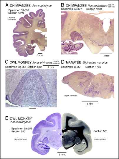
On the web site, viewers can see and download photographs of brains of over 100 different species of mammals (including humans) representing 17 mammalian orders. Also available are examples of stained sections (see images below) from a wide variety of brains of special interest, including series of sections from brains of humans, chimpanzee, mandrill, rhesus monkey, owl monkey, Atlantic bottle-nose dolphin, Florida manatee, California sea lion, African lion, polar bear, yellow mongoose (mierkat), short-tailed weasel, llama, domestic pig and cow.
 |
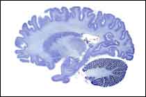 |
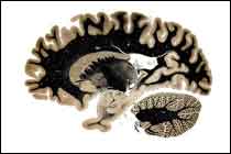
Section of human brain using fiber stain. |
Section of human brain using cell stain. |
Copyright National Museum of Health and Medicine at the Armed Forces Institute of Pathology, Michigan State University, and the University of Wisconsin. Reproduced with permission from the Comparative Mammalian Brain Collection. |
|
Along with the images of brains and sections of brains, there are searchable databases of information about the specimens, including body and brain measures, numbers and planes of sections, and other data of interest. These databases can be downloaded in several database formats. In addition, databases listing the contents of other comparative brain collections in Europe and America are available on the site.
Also on the site is an illustrated discussion of research in Paleoneurology, the study of fossil brains of extinct animals, by Dr. Harry Jerison.
Under development at a satellite testing site are more ambitious atlases of fully labeled brain sections. The atlas for the brain of the sheep is most developed at this time. All atlases can be seen, as they develop, at http://www.msu.edu/user/brains/atlases.
The generation of these web sites has been supported by The Division of Integrative Biology and Neuroscience of the National Science Foundation. Over the decades, preparation of the specimens has been supported by the National Science Foundation and the National Institutes of Health.
Contributed by:
Wally Welker, University of Wisconsin
<[email protected]>
John Irwin Johnson, Michigan State University,
<[email protected]> and
Adrianne Noe, National Museum of Health and Medicine
<[email protected]>
DOI: 10.1045/october2001-featured.collection